3D Cell Culture

3D cell cultures are driving new scientific breakthroughs in basic research, drug discovery and personalized medicine applications. High throughput, automated liquid handling solutions help researchers to work more effectively with challenging reagents and precious cells, to unlock the technology’s full potential.

How 3D cell cultures are advancing scientific knowledge and improving research outcomes

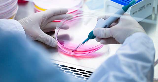
scientist-with-tissue-culture

In vitro cell cultures are crucial tools for modelling disease. Traditional 2D cell cultures - while simple to use- do not inform as effectively about biological processes as 3D cell cultures.(1) 3D cell cultures more accurately model human physiology and provide better insights for oncology, drug discovery, and personalized medicine applications. In this in-depth guide, we discuss the benefits of using 3D cell cultures, the different types of 3D cell cultures and their applications, challenges to adoption, and the role of automation in unlocking their full potential. 

Frequently asked questions

3D cell cultures can help us to bridge the gap between in vitro and in vivo research by:

  • improving efficiency and enhancing the data quality of pre-clinical assays
  • increasing the probability of success in understanding diseases such as cancer
  • generating more translatable drug responses for discovery and personalized medicine applications compared to 2D cell cultures
  • reducing reliance on animal testing

The rapid emergence of many techniques for culturing cells in 3D has led to inconsistent use of terminology, which now benefits from clarification. (2) Spheroids are simple, widely used 3D cell culture models, which form based on the tendency of adherent cells to aggregate – and may be generated from many different cell types. Organoids are more complex 3D aggregates, like miniaturized and simplified versions of an organ. They can be tissue or stem cell-derived with the ability to self-organize spatially and demonstrate organ-specific functionality. (3)

Further, several different cell culture formats support the growth of 3D cell cultures (4) encompassing:

  • Anchorage-independent (non-scaffold based): hanging drops, low attachment plates, micropatterned plates
  • Anchorage-dependent: biological hydrogels (e.g., Matrigel(R), collagen) or synthetic hydrogels
  • Organ-on-a-chip

It is important to note that there is no single gold standard technique. Instead, the optimal choice of 3D cell culture format depends on many factors, including:

  • Cell line and source (immortal, primary, iPSC, tissue explant)
  • The desired level of biological complexity/relevance to be modelled
  • Throughput requirements
  • Cost
  • Assay reproducibility

Anchorage-independent models are very scalable, economical, reproducible, and a popular choice for simple spheroid cultures. However, they lack interaction with an extracellular matrix, which is limiting for some applications. 

Anchorage-dependent cultures do not require specialized plates and are suitable to support both simple and complex 3D cell culture models. Animal-derived scaffolds, such as Matrigel ® or collagen, provide the mechanical and physical anchorage of cells found in vivo

However, some critics point out that the composition of animal-derived hydrogels can be variable between batches, and derivation from cancer cell cultures does not reflect healthy physiological levels of biological factors. On the other hand, synthetic scaffolds do not suffer from batch-to-batch variability but lack some of the attributes required to support normal cell adhesion, growth, and differentiation. (2)

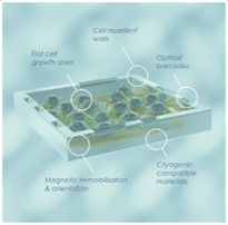
Organ-on-a-chip formats support the growth of complex and highly specialized 3D cell cultures that replicate the functionality and complexity of organs. They consist of microfabricated fluid channels that allow the growth of (embedded) cells and liquid perfusion channels.

There are now several commercial vendors of organ-on-a-chip technologies, though most designs are still in development and reside within academic institutions. The high cost of the chips has so far precluded their use from high-throughput screening applications, but they are increasingly adopted in R&D and smaller-scale screens.

Several fields now apply 3D cell cultures, including:

  • basic research
  • drug discovery
  • personalized medicine

As researchers have moved away from 2D cell cultures in favour of 3D cell cultures, a similar trend has emerged in the choice of cell line, with primary cells, induced pluripotent stem cells and co-cultures now favoured over immortal cell lines.

Genomics is playing an increasingly important role in 3D cell culture applications. Genome editing techniques such as CRISPR-Cas9 editing have given scientists the tools to investigate the effects of mutations on disease states.  In addition, next-generation sequencing (NGS) has allowed deep insight into the complex, often heterogeneous make-up of complex diseases such as cancer. 

3D cell cultures for basic research & disease modelling

3D cell cultures have been utilized extensively in basic research and disease modelling, including cancer. (5)

We now have compelling evidence from two decades of research that reveals the critical role of tumor microenvironment (TME) in cancer development and progression. In two-dimensional (2D) cell culture systems, cells are grown as monolayers on a flat solid surface, lacking cell-cell and cell-matrix interactions of native tumors. These 2D-cultured cells are stretched and undergo cytoskeletal rearrangements acquiring artificial polarity, which in turn causes aberrant gene and protein expression. In contrast, 3D culture systems offer the unique opportunity to culture cancer cells alone or with various cell types in a spatially relevant manner, encouraging cell-cell and cell-matrix interactions that closely mimic the native environment of tumors. These interactions cause the 3D-cultured cells to acquire morphological and cellular characteristics that reflect in vivo tumors.

In response to these advantages, researchers use tumour spheroids to study various mechanisms involved in cancer biology, including:

  • metabolic and hypoxia-induced changes
  • cancer cell invasion and migration
  • cancer stem cells
  • tumor microenvironment signalling crosstalk

Beyond oncology indications, 3D cell cultures have been utilized in other areas of basic research, including:

  • the study of neurodegenerative diseases (6)
  • development of in-vitro blood-brain barrier (7) models
  • pancreatic organoids, as potential models for pancreatic cancer and diabetes (8)
  • intestinal organoids to model cystic fibrosis (9)

3D cell cultures for drug discovery

Most potential drug candidates fail clinical trials (80%), and this figure is even higher in oncology. (10) The most common reason for failure is lack of efficacy, but a staggering 17% of phase 3 clinical trials fail due to safety concerns. (11)

3D cell cultures have become valuable tools at all stages of pre-clinical drug discovery, starting from disease modelling and target identification and verification through to screening, lead selection, efficacy, and safety assessment. Researchers often grapple with complex trade-offs between model predictive strength and cost, ease of setup, and throughput capability. Owing to the cost, complexity and difficulty in automating 3D cell culture models, drug screening is conducted on a modest scale, usually not exceeding 100,000 compounds. Indeed, screening is not yet routinely performed in 3D cell culture, but hits may be validated for efficacy and toxicity using 3D models. Working with a smaller number of compounds may permit the use of more advanced models or organ-on-chip technologies.

Within the drug development process, compounds undergo rigorous safety testing before entering the clinical stage.  Pre-clinical assessment of safety comprises in vitro testing (biochemical and traditional 2D cell culture assays), followed by animal testing.

3D cell cultures are not yet at a stage where they could replace toxicity testing in animals, but considering the high cost, ethical concerns and physiological differences between animals and humans, they may develop into promising alternatives. This  is enhanced by the development of new 3D cell culture formats that test for hepatotoxicity (12) and cardiotoxicity (13).  However, there is insufficient evidence to understand whether these approaches could improve upon the physiological relevance of animal testing. Or, on the other hand, whether there still remains a lack of complexity, for example to  account for the role of the vasculature or the immune system. (13)

Despite the  numerous challenges associated with implementing 3D cell cultures for drug discovery applications and high throughput screening applications in particular, the use of 3D cell cultures, together with better cell models such as stem cells and primary cells, could allow greater predictability of efficacy and toxicity in humans before drugs move into clinical trials, which, in turn, would lower the attrition rate of new molecular medicines under development.

3D cell cultures for personalized medicine

Molecular targeted therapeutics continue to improve the outcomes for many cancer patients. It is known that there is significant tumour heterogeneity among patients, as well as heterogeneity within a single patient tumour. This limits the efficacy of targeted therapies to specific patient subtypes and contributes to relapse.(14)

Genetic testing of tumours is already commonplace in the clinic to identify cancer subtypes and guide targeted therapies. However, the large number of genetic and epigenetic changes in many cancers and our lack of genotype-phenotype relationship understanding suggest that complementary strategies are needed to guide treatment.(14)  

Patient-derived organoids retain the heterogeneity of the original tumor but allow several different drugs or drug combinations to be tested for efficacy in vitro. Drug screens on patient-derived organoids are not yet fully validated as predictive biomarkers in the clinic. Therefore, more research and development are needed to standardize patient-derived organoids and refine the models. The creation of cancer biobanks that provide access to relevant cells will help to advance this goal. In the future, we may see patient-derived organoids and genetic testing as complementary tools to guide personalized medicine approaches. This promising development looks set to increase the efficacy of treatments and minimize toxic side effects.(15

3D cell culture techniques are used within pharmaceutical and academic settings and increasingly applied in personalized medicine.  With several commercial vendors of 3D cell culture plates and chips, specialized cell lines, and better biobank access, techniques are now becoming available to a wider audience.  Within the pharmaceutical sector, oncology remains the most common application of these methods, and in academia, they are applied across a wide range of research fields.

The barriers to adoption of 3D cell cultures fall into two broad areas (3):

  • Scientific validation of 3D cell culture models and quantification of their predictive power 
  • Process and technical challenge associated with standardizing assays, increasing throughput, developing analysis pipelines, and decreasing cost

Validation of 3D cell culture models

Numerous studies have shown that 2D and 3D cell cultures generate different drug responses, and it is not yet known how precisely 3D cell cultures will represent disease biology. Predictive values for both 2D and 3D cell culture models would be useful to determine the phenotypic footprint of successful interventions. Until such data are available, 3D cell cultures will remain complementary to 2D cell cultures and biochemical assays. While we may see gold standard phenotypic models emerge in time, there is currently no scientific consensus. Regulatory issues will need to be resolved, and with a renaissance of phenotypic screening approaches, target deconvolution will be critical in securing regulatory approval.  3D cell culture toxicology assays cannot yet replace animal testing. Nevertheless, there is potential value in their application as a supportive approach to minimize animal use by weeding out compounds with poor toxicology profiles earlier in the development lifecycle.

Cost

3D cell culture applications are significantly more expensive than 2D cell culture applications due to the need for specialized cells, reagents (basement membrane extracts) and consumables (specialized plates or chips).

Standardization and reproducibility

The high degree of biological variance in starting material and lack of standardized methods can lead to issues in assay reproducibility.

Achieving throughput can be challenging as sensitive cells and temperature-sensitive reagents are not straightforwardly amenable to automation. While simple spheroid assays in anchorage-independent culture formats are relatively easy to automate, this process gets much more difficult for organoids, relying on temperature-sensitive materials like Matrigel ® or organ-on-chip architectures. Setting up organoid cultures requires precise liquid handling with a high degree of positional accuracy and temperature control. High-throughput culture formats are still, for the most part, limited to 96/384 well plate footprint.

Lack of materials is a consistent issue, since primary or patient-derived cells are generally available only in limited numbers. Growing enough cells for a screen can take weeks to months.

Analysis pipelines are also problematic. While it is possible to determine some simple whole-well assay readouts (e.g., ATP quantification), the most comprehensive insights come from multiparametric phenotypic analyses to exploit the complex cellular response patterns to classify compound effects. These require high-content imaging and analysis pipelines, niche skill sets, and sufficient time to execute, limiting compatibility with some 3D cell culture formats.(2)

Achieving automation for handling organoids has been historically challenging for several reasons, including a limited supply of cells to support these models, prohibitive dead volumes on traditional liquid handlers and dispensers, as well as difficulties in handling viscous basement membrane extracts and setting up highly reproducible 3D cell cultures.(16) Given the modest throughput of many 3D cell culture applications, hand pipetting remains the gold standard in many laboratories.

Benchtop dispensers based on positive displacement technology offer the opportunity to bring significant process efficiencies to 3D cell culture setup and to standardize workflows for more reliable data.

Positive displacement dispensers are based on a piston in direct contact with the liquid inside the dispense syringe. With no air gaps or system liquid, this method is ideal to handle viscous materials like Corning Matrigel®  with high accuracy and precision. (17)

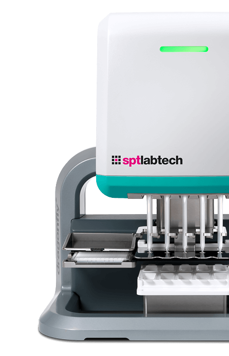
Systems like the SPT Labtech dragonfly discovery can achieve accurate dispensing starting from only 200 nL, enabling effective assay miniaturization. This not only decreases the cost of reagents and specialized cells but is also highly attractive when the starting material (e.g., patient-derived cells) is scarce. Combined with a low-dead volume of only 30 uL, it allows users to set up more replicates than would be possible by hand pipetting or traditional dispensing. Speed and temperature control are essential to handle ECM preparations such as Matrigel ® or Cultrex. Non-contact dispensing is similarly well-suited from a speed perspective, as a single syringe can rapidly (<1 min) fill all wells of a 96 well plate before the materials have had time to warm up to polymerize. Additional passive cooling modules can chill reagents on deck if multiplate dispensing is required. 

Automation allows users to standardize 3D cell culture setup, such that the only assay variables are the targeted interventions (drugs, genomic perturbations, RNA knockdown) and the heterogeneity of the input cell population. Given the need to implement HCA analysis pipelines, having a dispenser that can dispense in precise, predetermined locations of a plate or chip is highly advantageous as it cuts down on the imaging area to be covered by HCA. This is a useful feature for 2D cell culture phenotypic analyses and becomes even more critical in 3D where a z-stack of images may be required.

While the setup of 3D cell cultures can be greatly simplified using a benchtop dispenser such as dragonfly discovery, users still need to consider culture maintenance, e.g., exchanging cell culture media for long term culture. Organoid cultures in ECM are very fragile, and standard plate washer settings are too harsh and imprecise to remove media. Liquid handlers can be an alternative; however, they do rely on the use of single-use pipette tips, which can get expensive. Some researchers favour the use of magnetic beads inside the organoid, which allows transfer of the organoid to a safe position on the plate while media are being aspirated or replenished.

3D cell culture applications are ever-growing in popularity, and we expect this trend to continue in the coming years. (18) At present, there is still a trade-off between 3D cell culture model strength and cost, throughput, and availability. Automation can help to significantly lower the barriers to adoption, so researchers can choose the model that best answers their scientific question.

We anticipate an uptick in adoption of spheroid and organoid models for drug discovery applications (including screening) and the entry of translational medicine applications to clinical practice in the not-too-distant future. One of the great strengths of 3D cell cultures is the multiparametric readouts afforded by phenotypic analysis. Researchers already utilize genomic perturbations (CRISPR-Cas 9/RNAi) to model and understand disease biology. We expect to see NGS techniques used more and more frequently in combination with 3D cell culture techniques.

As scientists continue to develop new techniques and technologies to optimize 3D cell culture workflows, the future for research in this area looks bright.

  • Corrò, C., Novellasdemunt, L. and Li, V., 2020. A brief history of organoids. American Journal of Physiology-Cell Physiology, 319(1), pp.C151-C165.
  • Booij, T., Price, L. and Danen, E., 2019. 3D Cell-Based Assays for Drug Screens: Challenges in Imaging, Image Analysis, and High-Content Analysis. SLAS DISCOVERY: Advancing the Science of Drug Discovery, 24(6), pp.615-627.
  • Fang, Y. and Eglen, R., 2017. Three-Dimensional Cell Cultures in Drug Discovery and Development. SLAS DISCOVERY: Advancing the Science of Drug Discovery, 22(5), pp.456-472.
  • Langhans, S., 2018. Three-Dimensional in Vitro Cell Culture Models in Drug Discovery and Drug Repositioning. Frontiers in Pharmacology, 9.
  • Nath, S. and Devi, G., 2016. Three-dimensional culture systems in cancer research: Focus on tumor spheroid model. Pharmacology & Therapeutics, 163, pp.94-108.
  • Korhonen, P., Malm, T. and White, A., 2018. 3D human brain cell models: New frontiers in disease understanding and drug discovery for neurodegenerative diseases. Neurochemistry International, 120, pp.191-199.
  • Campisi, M., Shin, Y., Osaki, T., Hajal, C., Chiono, V. and Kamm, R., 2018. 3D self-organized microvascular model of the human blood-brain barrier with endothelial cells, pericytes and astrocytes. Biomaterials, 180, pp.117-129.
  • BMC Series blog. 2021. Human Pancreas Organoids: A step closer to understanding biology & treating disease - BMC Series blog. [online] Available at: <https://blogs.biomedcentral.com/bmcseriesblog/2020/02/26/human-pancreas-organoids-a-step-closer-to-understanding-biology-treating-disease/> [Accessed 26 May 2021].
  • Noordhoek, J., Gulmans, V., van der Ent, K. and Beekman, J., 2016. Intestinal organoids and personalized medicine in cystic fibrosis. Current Opinion in Pulmonary Medicine, 22(6), pp.610-616.
  •  Tanabe, L., 2020. Improving Drug Discovery with Organoids. [online] Biocompare. Available at: <https://www.biocompare.com/Editorial-Articles/566632-Improving-Drug-Discovery-with-Organoids/> [Accessed 26 May 2021].
  • Fogel D. B. (2018). Factors associated with clinical trials that fail and opportunities for improving the likelihood of success: A review. Contemporary clinical trials communications, 11, 156–164. https://doi.org/10.1016/j.conctc.2018.08.001
  • Sakabe, K., Takebe, T. and Asai, A. (2020), Organoid Medicine in Hepatology. Clinical Liver Disease, 15: 3-8. https://doi.org/10.1002/cld.855
  • Zuppinger, C. (2019). 3D Cardiac Cell Culture: A Critical Review of Current Technologies and Applications. Front. Cardiovasc. Med..
  • Li, Y., Tang, P., Cai, S., Peng, J. and Hua, G. (2020). Organoid based personalized medicine: from bench to bedside. Cell Regeneration. [online] Available at: https://www.ncbi.nlm.nih.gov/pmc/articles/PMC7603915/.
  • Driehuis, E., Kretzschmar, K. and Clevers, H., 2020. Establishment of patient-derived cancer organoids for drug screening applications. Nature Protocols, 15(10), pp.3380-3409. (5)
  • Yuhong Du, Xingnan Li, Qiankun Niu, Xiulei Mo, Min Qui, Tingxuan Ma, Calvin J Kuo, Haian Fu, Development of a miniaturized 3D organoid culture platform for ultra-high-throughput screening, Journal of Molecular Cell Biology, Volume 12, Issue 8, August 2020, Pages 630–643, https://doi.org/10.1093/jmcb/mjaa036
  • Sherman, John Shyu, Hung, Automation of Forskolin-induced Swelling Assay of Human Intestinal Organoids
  • King, D., 2020. A Look Towards the Future of 3D Cell Culture – A panel discussion. [online] Cell Culture Dish. Available at: <https://cellculturedish.com/category/general/> [Accessed 26 May 2021].

Enabling organoid research
through collaboration

In collaboration with Corning Life Sciences we combined the liquid-handling power of SPT Labtech’s dragonfly discovery and Corning’s Matrigel™ matrix for organoid culture, to automate a forskolin-induced swelling assay of human intestinal organoids. The team was able to effectively dispense 3 µL droplets of human intestinal organoids suspended in Corning Matrigel™ matrix for organoid culture into 96-well microplates.

The low volume/low dead volume dispense capabilities of dragonfly discovery allowed the user to make the most of precious resources, and enabled assays that would have otherwise been prohibitive from a cost or cell availability point of view.

Learn more about the project by reading our Q&A with Hilary Sherman, Senior Applications Scientist at Corning.

Anne-Hammerstein

Related products

With a focus on liquid handling, sample preparation and sample
management, our expert teams create state-of-the-art solutions
that scientists and researchers can trust.

dragonfly® discovery

dragonfly discovery enables innovative, low volume reagent dispensing across a broad range of applications including genomics​, assay development, and 3D cell cultures. Highly repeatable and accurate low volume dispensing, rapid large volume dispensing, and ultra-low dead volumes allow researchers to minimize reagent costs, save time, and standardize assays for superior quality data.

Explore dragonfly discovery
dragonfly-product-category---product-dfdiscovery